

Long overdue, we now have gorgeous digital (and printable) gift certificates for those who wish to give the gift of healthy breast (or other body) imaging to their loved one(s). There are no expiration dates for these gift certificates so your investment cannot be lost to time!
ABNORMAL Left Breast REVERSED Plus "Significant Improvement" in the Estrogen Pattern!!!
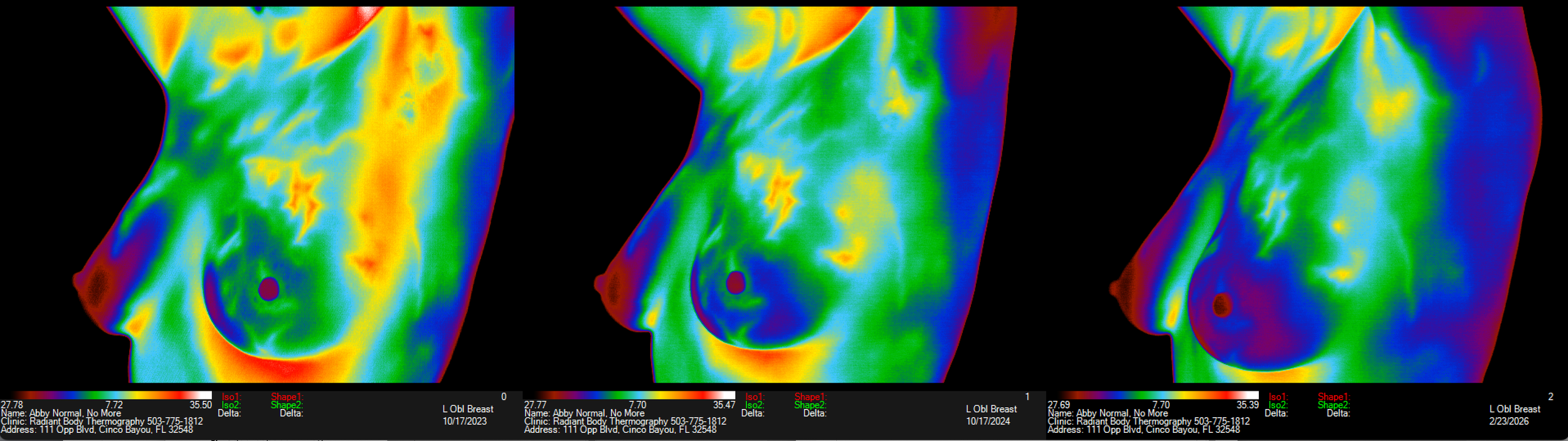
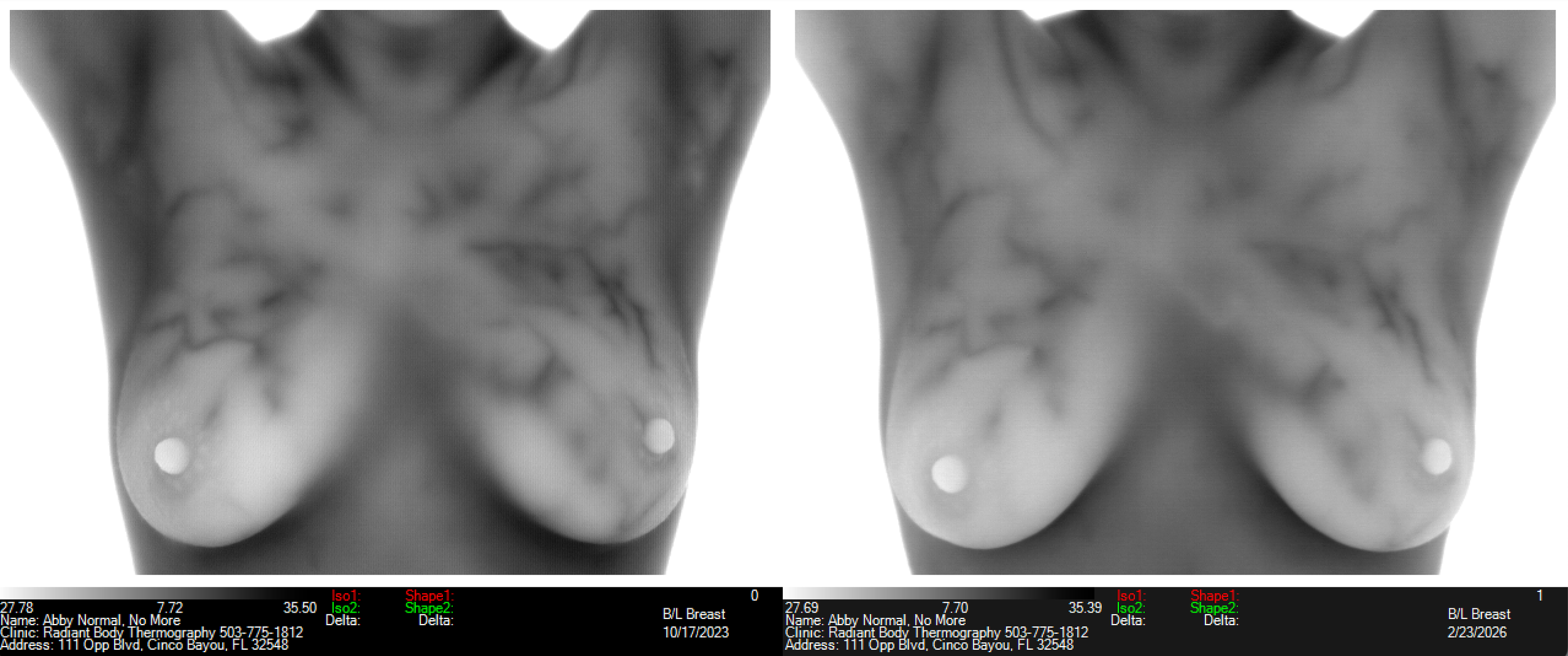
This client had an abnormal left breast for several years! All images of left breast below are “abnormal” (TH4) except the ones on the far right of each group with date of Feb 2026. It wasn’t getting worse but wasn’t getting better either, plus she had had the pattern suggesting estrogen dominance for all those years too, going back to 2018!
She moved to a much more southern latitude (Texas from Oregon in 2022?) plus she had a mercury amalgam tooth filling removed a few months before the last imaging. Says she doesn’t crave or eat nearly as much sugar as she did previously. Could this be due to more sun exposure which is balancing her hormones naturally? She was never interested in using progesterone cream.
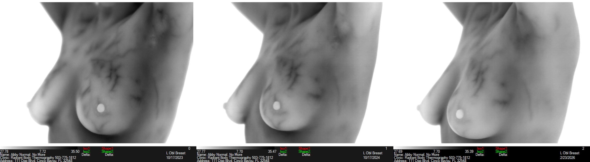
Lighter whiter breast as the estrogen pattern lessens - far right image shows “significant improvement”

Black is the coldest color (left end) and white (right end) is the hottest hue with this color scale.
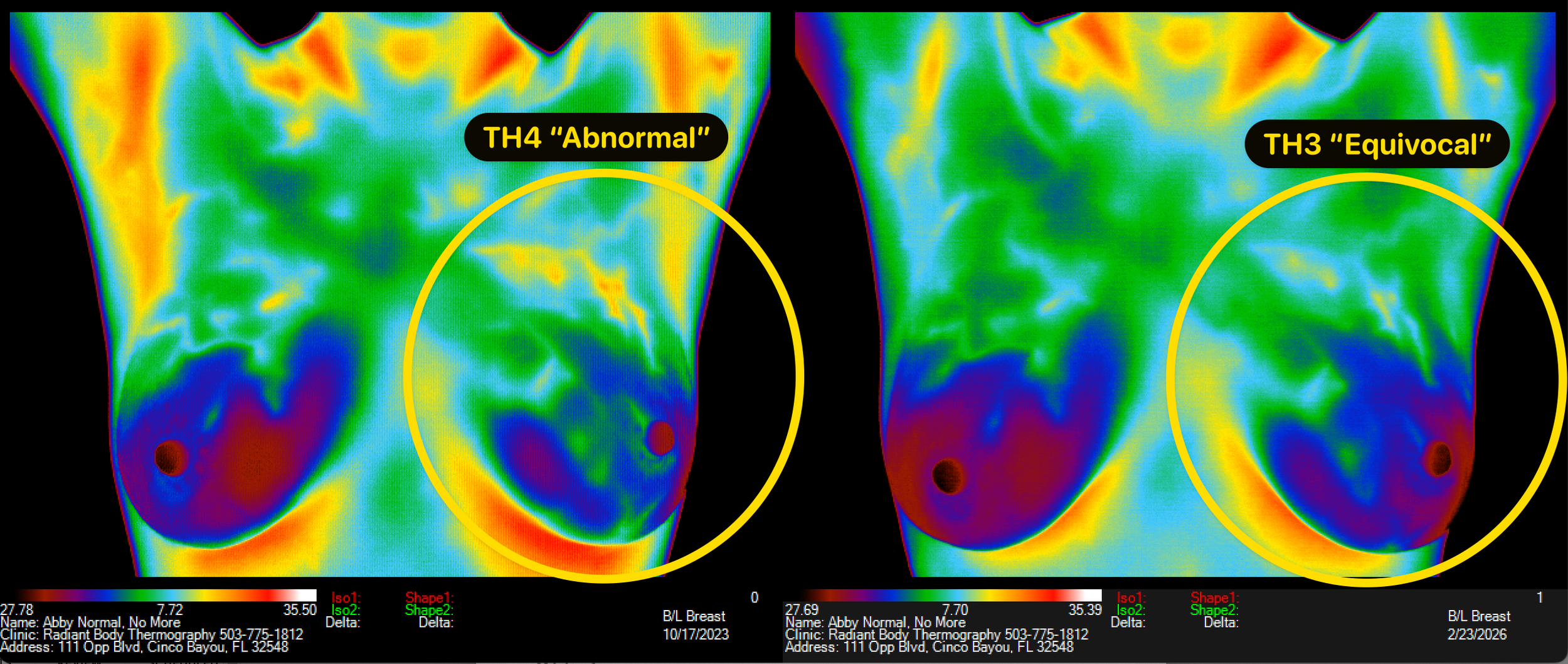
Clearly more symmetry in the far right image - she’s no longer losing her light/heat in her left breast!
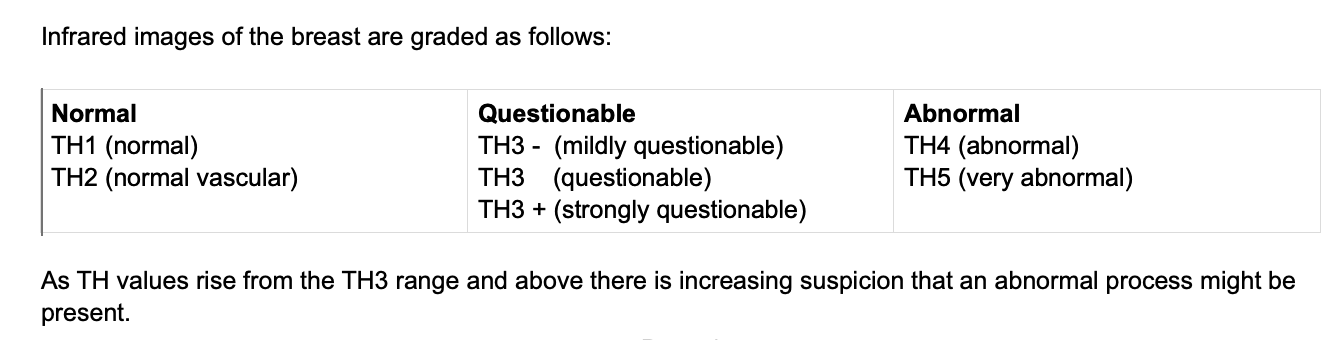
EQUIVOCAL is the medical term for TH3 Group which means not normal but not abnormal either, 95% probability of not being pathology/disease, yet the group of women who were involved in designing Dr. Amalu’s reports did not like the word “equivocal” because they’d not heard of it so “Questionable” was substituted. Not a great choice in my opinion as the technician because gets people unnecessarily excited.
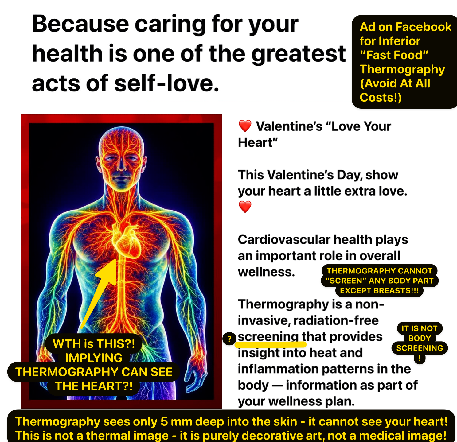
Recognizing Inferior "Fast Food" Thermography - Example of Misleading Graphics and Wording - FB Valentine Day Ad 🙈
A client sent me a Valentine Day ad that is a perfect example of a very deceptive ad for thermography implying that thermography can evaluate the whole cardiovascular system including the heart! This ad is heart-breaking, not heart healthy. See my notes in yellow writing.
Scroll down on this page to Thermography Myths. The very first one addresses the false claim you are seeing in this ad and there are several more worth your time if you are unsure about this technology’s capabilities.
Breasts are especially suitable for screening because they hang outside the body, are supposed to be cooler, so any abnormal heat “jumps out”. In an abdomen that is warm by nature because it is the core of heat, a disease could be very advanced before enough heat would exist to not be hidden by the normal heat, putting a person in huge danger to depend upon a thermal scan for internal organ screening. Thyroid is close enough to the surface that we do see stress signals but that gives no indication if it is hypo or hyper, just that there is stress. Better to use a ten-panel thyroid test for detail.
Screening requires/implies that we find early disease signals in an otherwise healthy person. Breasts are the only body part that thermography can screen (see early signals of disease). Anyone claiming to screen your heart or any other abdominal organ with thermography may as well be holding up a big red flag with “snake oil” written on it.
These FAQ’s from Dr. Amalu’s site, breast thermography.com, give one an accurate quick overview of clinical thermography’s amazing capabilities as well as its limitations. It is incredibly unethical to oversell thermography’s capabilities as SCREENING for any body part other than BREASTS. It cannot give information about gut issues either unfortunately.
Thermal Images Used To Sell Products, Pills, Potions, & Lotions - Which Images Can Be Believed & Which Are Potential Red Flags 🚩 Scam Alert🚨 Do Not Get Sucked In
Please do not be fooled by before and after “improvements” using thermal images if there are no detailed numbers on the image showing the SETTINGS USED. They must be the same, or very, very similar, if before and after images are to have a chance of being believed. This is your assurance that the seller is comparing apples to apples. Leaving these numbers off is so unprofessional because it is super easy to NOT CROP THEM OFF!
It is super easy to create “before & after” fakes instantly in a few clicks. Without the settings visible that presumable relate back to real date and time stamps and setting for SPAN and LEVEL, these images cannot and must not be believed.
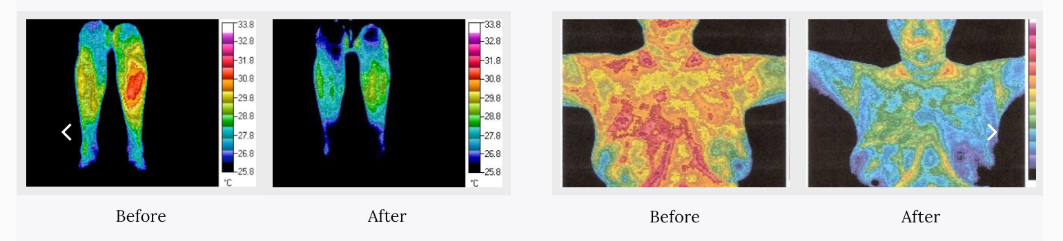
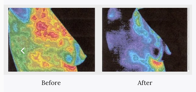
These may be a great products - the thermal images imply that a gadget (above images) and a pill (below images) reverse breast disease - why not publish the settings? Without the numbers, it’s not believable to anyone with skill in the thermal imaging industry who knows how easy it is to apply colors that may have nothing to do with reality.
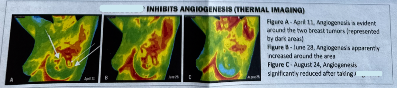
Proof using thermal images needs to show the settings even if too small to read because zooming in will reveal them. See how similar the settings are in images below from one to the next? This is your assurance that you are not getting sold a bill of goods.
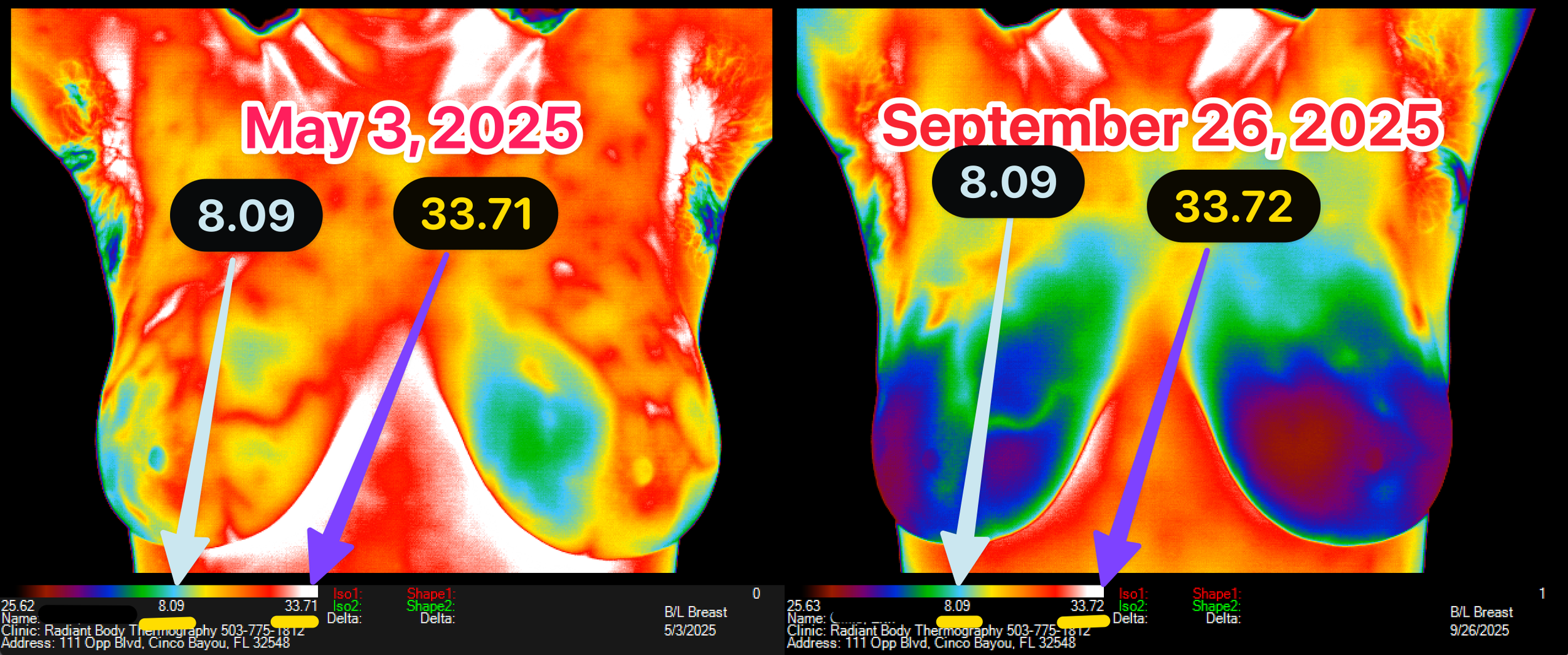
Zoom into those numbers and make sure they are same or similar; if they are missing, ask for uncropped images before sinking your money into products or pills using thermal images as “proof”.
Vaginal Support Moisture Proper pH Product - Natural Ingredients- Highly Recommended by RBT Client - Money Back Guarantee
According to a longtime client, there are lots of products for vaginal dryness, itching, pH imbalances and other conditions like Lichen Sclerosus that do not work and are a waste of money she says. But this product she loves. They make suppositories too which she also uses.
Neueve’s website claims to be saving marriages and offers a money-back guarantee. Formulated by the doctor selling it direct.

Causes of Estrogen Dominance (Exogenous and Endogenous Sources)
Exogenous Estrogen Sources
Contraceptives and HRT
Phytoestrogens (soy, flax, black cohosh, chaste berry, etc.)
Xenoestrogens
Sunscreens
Makeup, shampoos, face/body washes, lotions, deodorants – parabens
Plastics – BPA, phthalates, DEHP, PCBs
Building supplies – PCBs
Insecticides – atrazine, dieldrin, endosulfan
Herbicides – glyphosate (Roundup)
Feminine hygiene products and toilet paper
Animal protein sources
Beef, pork, chicken – growth hormone, estrogen.
Farmed fish – soy feed
Nutrient deficiency – B vitamins (B7 folate, B6, B12)
Enzyme production for use in estrogen clearance/detox
Magnesium – Estrogen detoxification
Over-consumption of alcohol - Slows or decreases the oxidation of estrogen in the liver (clearance)
Endogenous Estrogen Sources
Ovarian overproduction of estrogen
Corpus luteum underproduction of progesterone (especially in menopause)
Overproduction of estrogen in the breast (10-50 times higher than can be found in the blood)
Hepatic estrogen clearance dysfunction
Conversion dysfunction (DHEA, testosterone, etc.)
Cortisol interference due to increased adrenal output
Hypothyroidism
Gut dysbiosis - bacterial imbalance decreases an enzyme needed for conversion to the form of estrogen needed for elimination.
Genetic dysfunction
Methylation (carbon with 3 hydrogens is added to another compound) - common chemical process in the body. Vital for proper DNA function – allows engagement or disengagement from certain genes. If methylation is impaired, it can cause a host of problems.
Gene MTHFR responsible for making the enzyme that regulates the metabolism of folate (Vitamin B7)
Gene COMT - responsible for making the enzyme that regulates the levels of estrogen.
List provided by Dr. William C. Amalu, Founder of Eagle Institute of Clinical Thermology and author of chapter 25, “Infrared Imaging of the Breast” in the Biomedical Engineering Handbook: Medical Devices & Systems, Ed 3.
These images show increase in this client’s estrogen dominance after her first visit but by the third visit, only five months after second visit, she and her doctor reversed the signal and report reflected the images have “significant improvement” in pattern representing estrogen dominance. (see her story here.)
TherMOMetry is Not Thermography and Should Not Be Trusted As Screening Device
Native Medicine Doctor with Tens of Thousands of Followers on InstaPuke Making Unproven Claims About What He's Calling Thermography That is Actually Thermometry!
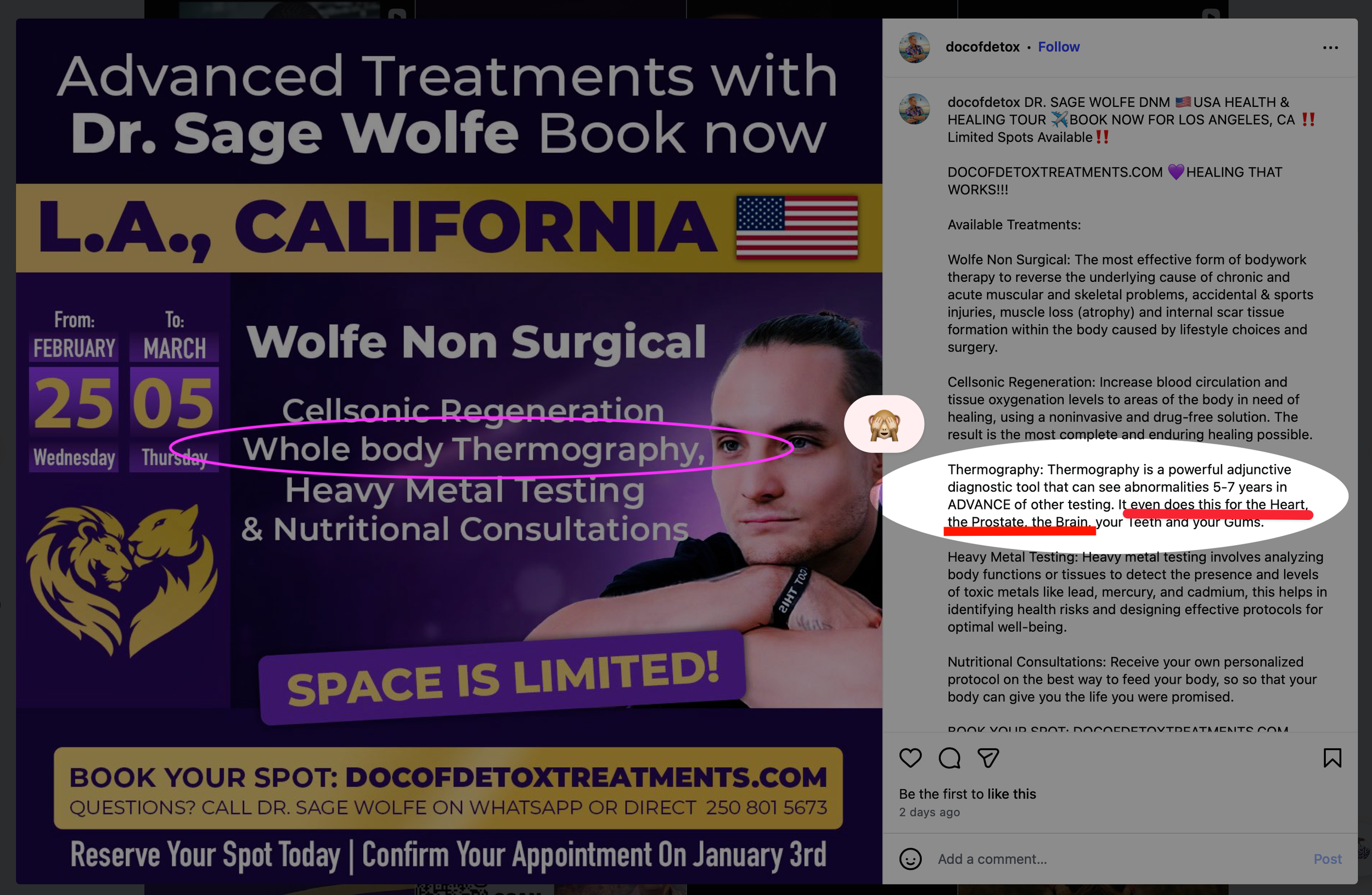
☝️MISLEADING CLAIMS insinuating he can accurately screen with his tool - clever wording but highly unethical given limitations👇
“Thermography is a powerful adjunctive diagnostic tool that can see abnormalities 5-7 years in ADVANCE of other testing. It even does this for the Heart, the Prostate, the Brain, your Teeth and your Gums.”
TOTAL BS!
Someone needs to ask Mr. Sage for the clinical trials showing efficacy of his claims. Are they claiming clinical trials that are of thermography yet using a wand or touch device? There are no clinical trials for touch devices that we know of so if they produce clinical trials for thermography which uses cameras, beware. Thermometry is a wand or tool that touches the body at different points - there is no “graph” or picture produced as in thermography. One such device can be seen at www.alfathermo.com
A search turns up the doctor’s Facebook page with video of a woman describing her “whole body thermography” (actually whole body THERMOMETRY is what she received) experience as a test of 160 points on her body.
Real whole body thermography measures the temperature in each pixel of the image down to 1/100 of a degree, registering many thousands of computer analyzed temperatures in a single image.
Please see Page 6, paragraph 3.3 of IACT Standards and Guidelines here to learn about the limitations of Dr. Sage’s device compared to a thermography camera system like we use.
If you read the entire document, you will learn that quality medical infrared THERMOGRAPHY is a fabulous way to screen for only one kind of cancer: it excels at detecting early signs of breast abnormality because breasts hang outside the body and are naturally cooler making extra heat very obvious. Not so for brain, heart or prostate as this so called doctor claims - nor is his thermometry device! And it certainly cannot produce comprehensive information about breast health measuring only a few points on them! Touching the skin with this instrument or any touching of the skin at all affects temperatures and is inherently not as accurate as measuring temps with an infrared camera. This is an extremely cruel, misleading dangerous post, especially for those living with cancer who may have limited time or money or both when facing a rapidly growing tumor and are in need of accurate, timely information.
REGULATION THERMOMETRY has its place but there are no clinical trials that we can find showing that is useful for screening which is what is being suggested when the claim is seeing 5-7 years in advance! Let’s see the evidence.
There is a thermography trial that got such significant results for ability to predict that it will never be repeated because it would be unethical to not make efforts to intervene knowing what we know from the trial which is that with abnormal breast scores in which no disease is detectable by other imaging at the outset will develop pathology within five years if no intervention to reverse the risk is made.
THERMOGRAPHY, if high quality, can detect heat consistent with growing breast tumors and estrogen dominance, a heat pattern which often precedes breast cancer, and it is a great tool for visualizing unresolved pain in 99% of cases. But perhaps it’s greatest use of all is for cancer patients who need to monitor treatment for efficacy, in other words to know if treatment is working which will be obvious as the extra heat dissipates from the tumor site.
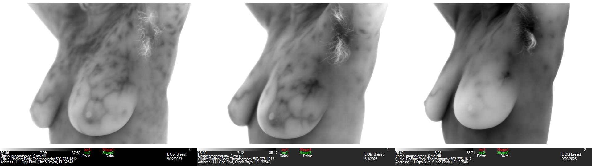
Risk-Lowering Power of Compounded Progesterone Capsule on Estrogen Dominance in Only 5 Months! Risk Reversal Clear as Day
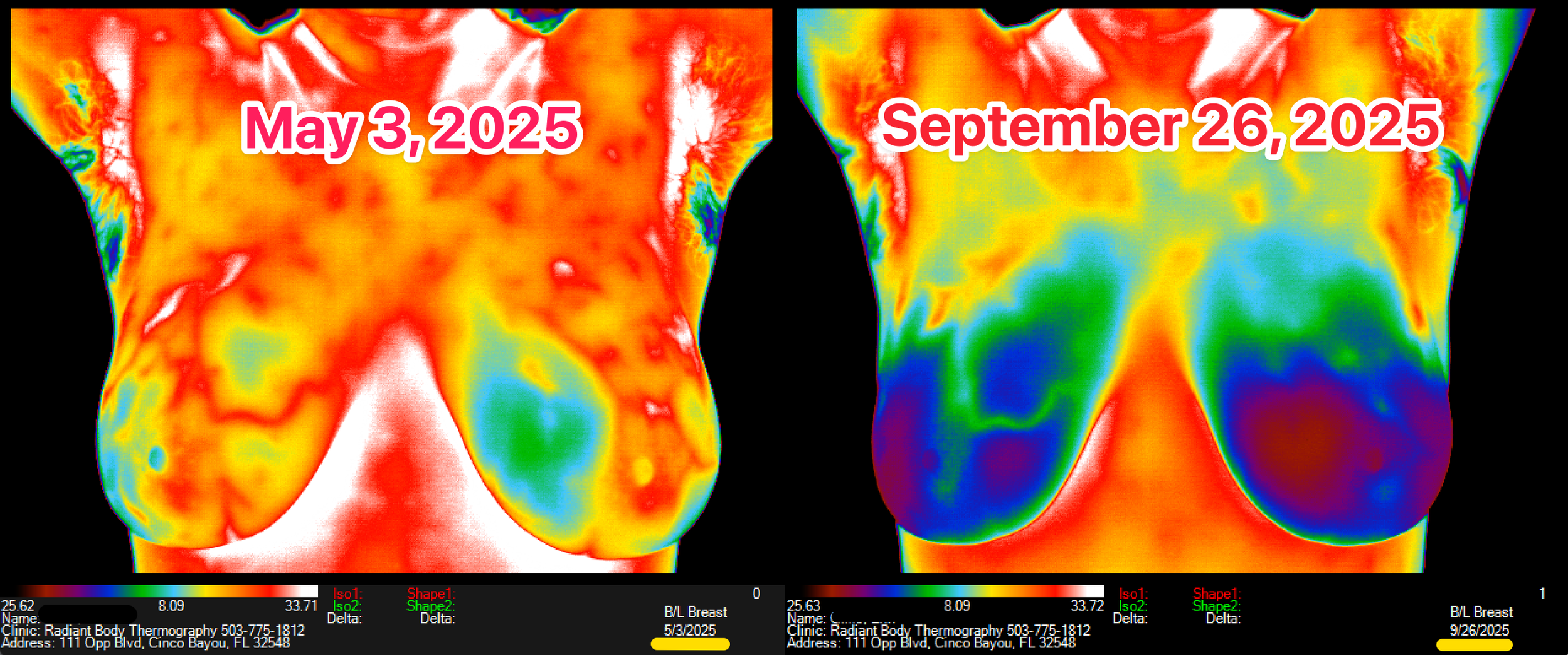
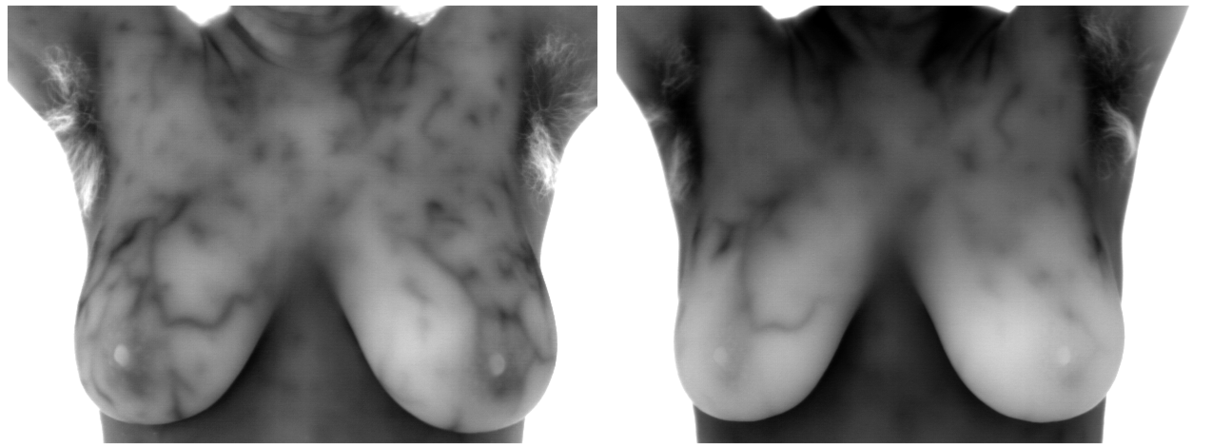
Black & White images show the ”significant improvement” in the pattern of estrogen dominance in approximately 5 months, plus she lowered her risk so much that her left breast got a better grade. Heat around a nipple can signal a problem and her left breast had heat encircling the warmer nipple.
This client’s story is fascinating and illustrates how important it can be to attend followup imaging plus how important to refrain from assuming that commercial formulations of bio-identical hormones that give undesired results are the end of the road.
Client’s first attempt at using progesterone to improve estrogen dominance symptoms was via Prometrium, a commercially available bio-identical brand. Unfortunately, 100 milligrams made her “feel like a zombie” so her doctor switched her to a compounded (personally mixed for her) progesterone capsule. No more zombie effect and she was able to increase to 200 mg. Black and white images (black is hot) show the powerful risk-lowering effects in only five months!
She felt so much better overall that she became more active, stronger and more fit, even shed a few pounds as you can see in the images!
Images above depict her second and third thermograms, below are all three. Her second imaging showed worsening in left breast, so she and her doctor, Kathryn Walker at Renu, developed a plan and executed it beautifully to reverse the trend! (Last we heard Walker is booked 9 months out but there are more brilliant practitioners - a client survey of who is/are your favorite doctor(s) is upcoming and we will publish results on this blog to give you more options!)
Below you see first session Sept 2023, second May 2025 and third Sept 2025. Her left breast increased in signal around the nipple by the second imaging and in the dark area leading up to armpit increasing her left breast’s grade between visits 1 & 2 but by visit 3, she had reversed the trend, grade returned to 2023 grade and estrogen dominance pattern is visibly even better than where she began in 2023 after only five months of getting hormones dialed in!
What contributes to Estrogen Dominance? Sources, Exogenous & Endogenous Ones
This page has a whole section of linked studies showing power of bio identical progesterone in prevention of breast cancer as well as its role in reversing estrogen dominance.
Progress Toward Truth in Screening! Density Must be Announced to Patient by Mammographers - But WHY? The Answer Will Shock You.
After decades of pleading by women (and the men who loved them) who were failed by mammography due to its reduced ability to see into their dense breasts, it is now mandatory that imaging centers performing mammagrams in every state must tell you if you have dense breasts.
The FDA updated its decades-old mammogram standards.
This is important because:
“Dense breast tissue is common in younger women. Dense breast tissue absorbs significantly more radiation during mammography compared with fatty breast tissue (1). This occurrence reduces the accuracy of mammography to detect breast cancer in women with dense breast tissue (2, 3). ”
What is buried between the lines is that dense breast tissue appears to raise one’s risk BECAUSE mammography is the screening tool, not because dense tissue is somehow abnormal or inferior tissue. To simplify, dense breast tissue is all the tissue minus the fat. The greater proportion of fat in the breast, the more likely the rays of radiation will pass through the tissue and into the plate beneath. Conversely, the higher proportion of non-fatty tissue, the greater the rate of absorption according to the American College of OBGYN, and also the greater likelihood of the appearance of interval cancers between mammograms for those with the greatest density (i.e., least amount of fatty breast tissue).
And if that wasn’t enough of a problem, cancer AND dense tissue both appear white on a mammogram, making the radiologist’s job extremely difficult; basically it’s like looking for a snowball in a snowstorm!
Which begs the question, why in the world would we choose a technology with reduced accuracy AND greater harm to women as the gold standard? Sadly it appears to be part of the system designed to protect the “geese” that lay the “golden eggs.”

Is the Breast Screening "Gold Standard" Mammogram Losing Its Shine?
A recent Medscape publication (a continuing education piece made into a quiz) about modern breast screening recommendations for clinicians called Raising the Bar on Breast Cancer Screening and Management sheds interesting light on the “gold standard” of screening: mammogram. Basically it concludes that if one wants to know what is truly happening, MRI is the best tool, though they do not mention counseling patients about the risk of the gadolinium contrast ending up in the brain nor the common allergy to the contrast agent. (Of course the Cinderella of Screening, Thermography, isn’t even invited to the conversation, possibly because it would be such a profit crushing blow in stopping treatment of lesions that are not actually cancerous and not destined to become cancer?).
Mammogram and ultrasound pretty much get thrown under the bus for the task of monitoring a suspicious lesion and for good reason in my opinion. Too many times for comfort, I’ve seen mammograms and ultrasounds come back “clean” when thermography is suggesting otherwise. One client getting asked for a biopsy, stating “I can see a little raised area where thermography is showing I have extra heat, may I have a biopsy?” Biopsy was refused for 3 days of her persistently requesting one so they finally gave her one and it turned out to be malignant! Then they said, “Oops! You need an MRI!”
MRI is also the tool most likely to see what Thermography sees as they both are able to visualize blood flow and blood flow in suspicious patterns can be indicative of angiogenesis, the formation of blood flow specifically for the purpose of feeding a tumor.
Multiple models, and some surprising risk factors get revealed when answers to the quizzes are shown as one completes them. Very educational for me! Hope you enjoy it too.
P.S. BTW, Dr. Amalu, interpreter of our images, is of course a huge fan of thermography combined with MRI for the very reasons stated above, i.e., these two are most capable of seeing blood flow which is sure to be present with a dangerous lesion.
Self-Proclaimed Estrogen Hormone "Expert" Wendy Sellens Gets Respectfully Corrected by Peers Which Becomes An Estrogen Lesson For All of Us

Wendy Sellens casts a dark shadow on the Father of Thermography, Dr. William Hobbins, M.D., quoting research that no one can find that he did, which according to her is that estrogen exists only in sex organs and she “knows” this because of tissue samples Hobbins supposedly took on all this other human tissue! ? What?!
Is it possible she wasn’t listening to Dr. Hobbins? (Want to assess whether she listens to her interviewers? Interviews below*)
Two people in the health field, Jay Feldman and Mike Fave, felt compelled to shine light on Wendy’s mistakes lest they mislead:
Ep. 125: Wendy Sellens on Estrogen: What She Got Wrong Nov 7, 2024
As a huge fan of Dr. William Hobbins’ work in thermography, it was beautiful watching complete strangers, Mike and Jay, come to his defense when he is six feet under and no longer able to speak for himself.
To their beautiful credit, Strong Sistas recently interviewed the two gentlemen that made the rebuttal to Wendy’s questionable claims, showing their ability to recognize truth and embrace it; those are here:
Why Modern Biohacking is Making You Sicker Not Stronger
&
Male Fertility is Falling Fast & Her'e’s Why (With Mike Fave). Estrogen comes up again of course in male fertility.


Mike and Jay call out by name all kinds of “medical experts” making claims that they feel are inaccurate with potential of harming others.
For example, they show, Dr. Ken Berry’s claims as highly questionable in Ken Berry’s Fructose Fearmongering, Glycation Myths, & ChatGPT Says We’re Wrong.

Jay and Mike also address AI’s deficiencies, particularly the risks when questions aren’t perfectly composed by someone that actually knows the correct answers, i.e., incorrect answers 🙈)!
Have you heard that AI has come to thermography? Beware of offices claiming superiority or higher accuracy because they utilize AI… it’s actually an old program out of India. Dr. Amalu can give you a thorough explanation of why it’s not ready for prime time.
*BELOW: NOT RECOMMENDED VIEWING, posted only in case you want to verify statements of Sellens.
Are you Actually Low in Estrogen? Jul 29, 2024
Too Much Estrogen or Not Enough? Aug 13, 2024
How Estrogen Affects Your Health . Aug 21, 2024
Hormone Masterclass: Estrogen Level 1 W/ Dr. Wendy Sellens Aug 28, 2024
Glyphosate (ingredient in ROUNDUP Weed Killer) INDUCES Breast Cancer Cells Growth
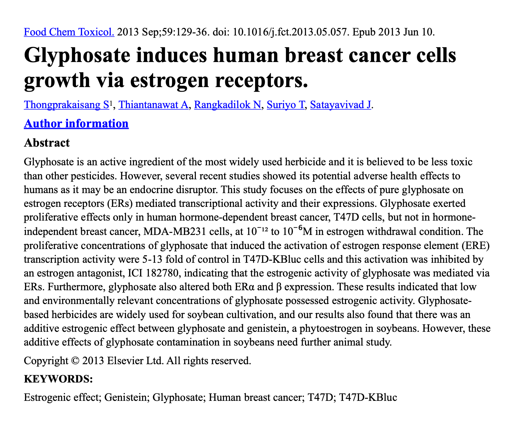
This is old but widely unknown news from the Food Chemical Toxicology “glyphosate contamination in soybeans” appears to be the biggest culprit but also many are still unaware of the big 5 crops that are genetically modified so that they will withstand multiple sprayings of Roundup. Try spraying it on your garden and you will see that it kills everything.
Big 5 GMO crops to Avoid:
soy
corn
canola
cottonseed
sugar beets
Sugar that is not labeled “organic” or “cane” will be from GMO sugar beets. Oils from soy, corn, canola, cottonseed are in so many things it will blow your mind. Also mind-blowing is that cancer patients are not typically warned about this nor is the hospital food free from glyphosate-containing foods nor is it advised in dietary advice from the hospital to avoid foods containing glyphosate.
Off the topic of breast cancer but important research on the subject and a possible second pathway of contributing to breast cancer is disruption of the detoxification process:
“Glyphosate chelates aluminum, allowing ingested aluminum to bypass the gut barrier.” (What about INJECTED aluminum!?) Glyphosate is linked to disruption of the gut-brain communication via disruption of the P450 cycle of the liver detoxification process, so the process by which a body would detox the chemical gets broken by the substance that needs to be detoxed. Dr. Stephanie Seneff has been warning us for at least two decades about this risk. She believes glyphosate is the biggest contributor to the escalating autism rates, and why symptoms can greatly improve with diet changes.
Lots of people are suffering from gut issues, bloating, gas, pain without having a clue as to its origin which could very well be glyphosate coming into their bodies.
It’s even in the air due to fuels being made with GMO corn that when burned aerosolizes the glyphosate according to experts. Even though advertised as “sustainable”, it’s probably best to avoid these fuels when possible, as well as the big five “body fuels” listed above.
The estrogenic effects of these crops may be one of the biggest contributors to the epidemic of estrogen dominance we see in men and women’s breasts.
Were you flagged with the estrogen dominance hormone pattern in your breasts? If you don’t know, contact us and we will look it up. Most thermography clinics typically do not report this pattern, so choose wisely if you are looking for thermography services. You want to receive not only this hormone pattern information but also you want to receive TH scores for each breast (TH1-TH5 are possible) and detail about location of findings.
United Breast Cancer Foundation Partially Reimburses For Thermography Expenses Consistently Per Client
Today I found out details about how one of my clients has been reimbursed for thermography imaging “3 or 4 times but not the full amount - varying amounts each time” after going through the UBCF’s Questionnaire & Application process. It’s 2 Steps: Questionnaire lets you know if you qualify to apply. Here is a list of the requirements (not on the list is that one must make a donation of $5 and my client says that solicitations for donations will arrive post grant receipt, so buyer beware.
Locations At A Glance; Where We Go & When + Scheduling and Event Detail Links

Click map to Schedule and see WHERE WE GO & WHEN! Imaging Radiant Bodies Across America ~ Bringing High Quality Thermographic Imaging as Close as Possible to as Many as Possible. Breast Screening at its finest and we also image full bodies - great for those who have unresolved pain issues.
Lake Havasu City, Arizona
Eureka, California
Sacramento, California
Boise, Idaho
Portland, Oregon - SE Cottage
Portland, Oregon, Westside - Bethany
Eugene, Oregon
Redmond, Oregon
Keizer, Oregon
Cannon Beach, Oregon
Ashland, Oregon
Roseburg, Oregon
Albany, Oregon
Vancouver, Washington (Brush Prairie)
Vancouver, Washington (Felida)
White Salmon, Washington
Woodland, Washington
2025 Autumn Tour of CENTRAL & WESTERN STATES! ~ 🚙🍁🇺🇸 ❣️Arizona, California, Colorado, Idaho, New Mexico, Oklahoma, Oregon, Washington

We are honored to once again take the reading skills of the country’s best and most experienced interpreting clinical thermologists at Eagle Institute of Thermology, Dr. William C. Amalu and his colleague, Dr. Valerie Quijano, to as many as possible by taking our thermography cameras on the road.
SEPTEMBER
Broken Arrow, Oklahoma (Tulsa suburb)
Longmont, Colorado
Boise, Idaho
Redmond, Oregon
White Salmon, Washington
Vancouver (Brush Prairie), Washington
Woodland, Washington
Vancouver (Felida), Washington
Cannon Beach, Oregon
Portland - West, Oregon
Portland - East, Oregon
OCTOBER
Keizer, Oregon
Albany, Oregon
Eugene, Oregon
Roseburg, Oregon
Ashland, Oregon
Eureka, California
Sacramento, California
Lake Havasu City, Arizona
Albuquerque, New Mexico
Littlefield, Texas
Oklahoma City, Oklahoma
You’ll receive private High Quality Infrared Imaging performed to the highest IACT Standards & Procedures by Candace Parmer, a certified thermographic technician using state-of-the-art cameras with over a decade of experience. Images are interpreted by world expert IACT Board Certified Thermologist, Dr. William C. Amalu, (author of Chapter 25, Infrared Imaging of the Breast in the Biomedical Engineering Handbook: Medical Devices and Systems, 3rd Edition) or his backup colleague.
Interpretation is included, no extra fees to speak to a person or doctor. Provided is a legitimate Medical Report that includes Findings, Digital Images, Recommendations, Guide (explains all possible grades, not just yours), all emailed via HIPAA compliant encrypted email 2-3 weeks after imaging, and 48 hr turnaround time is available for a fee for those who find waiting intolerable or who have medical reasons to have results sooner.
Tour Map - Imaging Radiant Bodies Across America❣️
😎 Summer Tour 🚙 📸 🇺🇸 Imaging Radiant Bodies in Florida, Texas & Alabama ❤️🔥 June, July & August ❣️
Dates and places have been finalized and links should all be working now 🥰 on the scheduling page and scheduling map.
Besides at our Florida HQ Clinic, we’re imaging in Pensacola, Panama City, Venice, and St. Augustine in June (and in January repeating those and adding Orlando back into the lineup).
Excited to reconnect with clients new and old in San Antonio (Bulverde area) and Austin area at these two fabulous spaces for imaging during mid July.
July & August we return to Gulf Shores & Gadsden Alabama where the sweetest people are found taking life in stride regardless of the heat or hurry around them.
When not on the road, you’ll find us imaging at our headquarters clinic in the western panhandle of Florida, Fort Walton Beach, right next door to Destin, “the world’s greatest fishing village” 🎣 🐈⬛.

🚙📸🇺🇸 Imaging Radiant Bodies Across America: Spring 2025 Tour ~ Dates & Places Here!

Click Map to Schedule from Map of all Locations We Image
In order of appearance we begin imaging Radiant Bodies on April 12th in Albuquerque NM, ending May 22nd in Tulsa, OK, and many stops in between including the Pacific Northwest!
(click links for more info about each event)
Albuquerque NM
April 12
Lake Havasu City AZ
April 13
Las Vegas NV
April 14
Eureka CA
April 16-17
Ashland OR
April 18-19
Roseburg OR
April 19-20
Eugene OR
April 20-21
Corvallis OR
April 21-23
Keizer OR
April 23-24
Cannon Beach OR
April 24-26
Vancouver WA
(Brush Prairie)
April 28
Vancouver WA
(Felida)
April 28-30
Portland OR
(Cottage on SE Crystal Spgs Blvd)
May 1-11
Portland OR
May 12
Washougal WA
May 13
White Salmon WA
May 13-15
Redmond OR
May 15-16
Boise ID
May 17
Golden CO
May 19
Littlefield TX
May 20
Tulsa OK
May 21-22
Beat the rush of fall’s breast cancer awareness season and take advantage of these spring imaging opportunities - more options, less time to get reports back, more chance of getting interpreted by Dr. Amalu instead of his backup, although YOU may request him specifically as the client patient at any time of year.

Lilacs in Full Bloom - April 11, 2025, one day shy of the April 12 Full Moon in Albuquerque, NM at Casas De Suenos Historic Inn, view from our Kachina Casita, our imaging studio when in ABQ
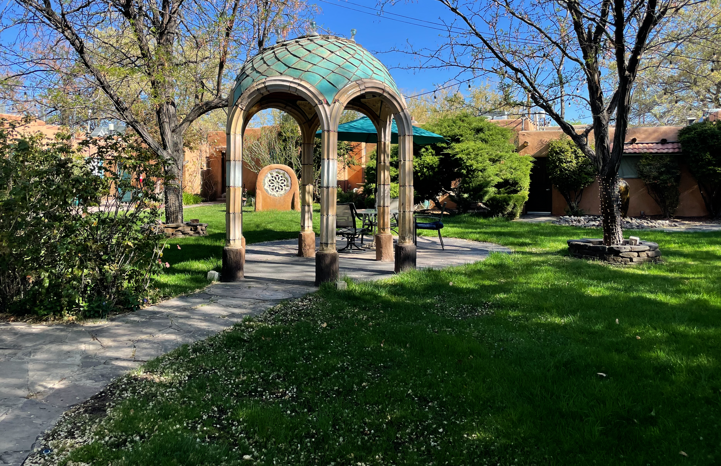
April 12 2025 Gardens at Casas de Suenos Historic Inn, Albuquerque, NM
🎉Radiant Body Thermography is Eleven Today! 🎂 Here's How We've Grown! 😎 Valentine Gift for Current Client Subscribers 💌
Radiant Body Thermography became a reality on Valentine’s Day 2014❣️


Improvements recently implemented:
📍 HIPAA-compliant self-scheduling for all locations from a map of 36 locations!
💄 Website makeover - organization plus lots of new photos are built into the design - photographs of cross-country road trips to life in Florida better reflect the mission to provide quality access to those we love.
💞 Online payments are possible but not required when reserving appointment so that those wishing to pay with cash or checks may still do so at time of service.

Beautiful Freak Snow on our driveway at Headquarters in Fort Walton Beach, Florida - 2025-01-22. 7:36 a.m.
5 Reasons Boutique Hotels And Homes Are Perfect for Clinical Thermograpic Imaging Sessions

Plaza Hotel, DT San Antonio, Texas
Boutique hotels give us complete control over temperature, drafts, and light making them ideal clinical thermography imaging studios
High end hotels with suites and short term rentals (Houses) are perfect in that there is plenty of space to do even full bodies and total control over temperature, light, drafts, privacy, all of which can be difficult to have in a clinic setting where others are working with different needs if each room doesn’t have its own temp controls and adequate space, window coverings, baffles, etc.
Some clients report being afraid to go into clinics so this scenario is avoided. Others appreciate the privacy, anonymity and atmosphere of hospitality in a high-end hotel or a clean cozy home.
At the inns, staff welcomes & caters to clients meeting clients so you are treated as a welcome guest along with any guests you wish to bring to your imaging session.
And a few clients want to book their next annual appointment on the spot. Clinics are often unable to plan a year in advance to host us but high-end hotels and homes or offices of dedicated fans 💞 do allow us to plan ahead to better accomplish our mission of increasing access to quality clinical thermography across the country.
Fast-Food Thermography is Not Your Friend - Insist Upon Quality Thermography - Avoid Disaster With These Tips
This short news brief published by Natural Awakenings written in 2022 gives several characteristics of fast-food vs. quality thermography although I omitted perhaps the most critical difference due to space limitations: a numerical TH (thermo-biological) Score (TH1-TH5) for each breast is given in quality reports, not in fast-food reports. While a TH score doesn’t guarantee quality, it drastically improves odds.