
Stay Radiant ~ Screen Safely!
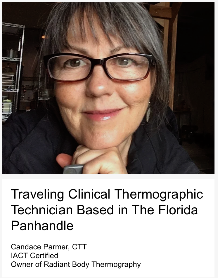
Quality Clinical Thermography ~ Breast Cancer Screening & Body Imaging for Radiant Bodies From Florida to the Pacific Northwest❣️
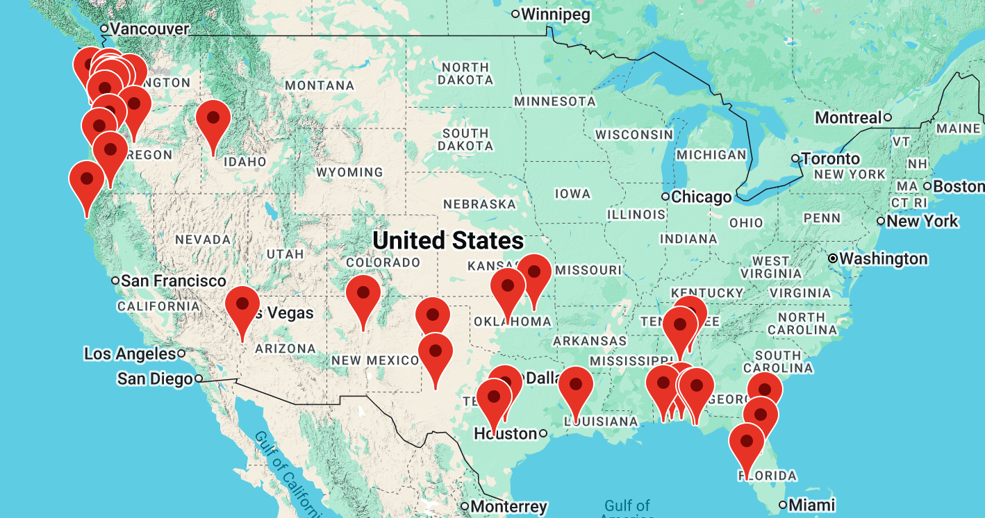
🚙 💞🇺🇸Radiant Body Thermography ~ Imaging Radiant Bodies Across America❣️
Say ‘Goodbye’ to inferior “fast-food” thermography because screening for breast cancer is serious business and
Your Life Matters!

Got Dense Breasts? No Problem! Thermal Breast Cancer Screening is Not Affected Negatively By Dense Breast Tissue 🥰
Thermography’s Sensitivity and Specificity are not lowered by breast density. What Are Your Breasts’ TH (thermobiological) Scores? Are breasts radiating a pattern suggestive of estrogen dominance that you may want to confirm & address with your clinician? What might your patterns be trying to tell you?
Our
SOLE FOCUS
is
THERMAL IMAGING
At Radiant Body Thermography, we sell no other products, pills, potions, lotions, oils or gadgets, and we are happy to GIVE you all the information and support that you need to incorporate this extremely accurate beacon of truth into your life
Mammography’s 85% sensitivity rating (on its best day in fatty breasts) is increased to 95% sensitivity by adding high resolution digital infrared imaging to a woman’s breast screening
Our Non Destructive Thermographic Breast Screening Includes Imaging & Assessment Via TH Scores, established by the IACT to Ensure Quality & Consistency
An abnormal infrared image is the single most important marker of high risk for developing breast cancer
~ All Ducts Lead to the Nipple ~
A Significantly Warmer Nipple On One Side is a Signal & Should Be Considered an Alert of Potential Pathology Especially if Progressively Getting Warmer
GREAT NEWS!
Most Breasts Receive TH Scores Giving Them 95% Odds That Pathology/Disease Is Not In Progress
Yes, we do full body thermography too but thermographic imaging cannot screen for cancer anywhere except breasts - breasts are naturally cool because they hang outside the body so abnormal heat pops right out in breasts. Your abdomen/core is naturally warmer allowing problems to hide from thermography too long to make it valuable as screening for cancer in the core of the body. Run from clinics claiming otherwise!
Your Body RADIATES
heat patterns
all the way to the lens of our camera!
Nothing is Beamed At or Into You Ever
100% SAFE
Equally effective
for all age groups
Zero Radiation
Zero Contact
Zero Pain
Zero Injections
Cruelty-free
Life-preserving
Ethical
Humane
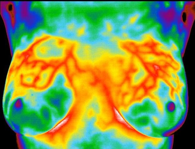
Quality breast thermography detects radiant heat signals and patterns from superficial blood vessels which can be consistent with expanding breast tumors with high metabolic rates (growing) and/or estrogen dominance.
High-quality detailed images and thermography reports are produced here, nothing like the dangerous "fast-food" thermography reports so common today.
Get The Test That Finds the Breast Cancers That Matter - the Hot Ones Destined to Progress, Not the Cold “Turtles” Going No Place Fast Enough To Matter (over-diagnosis) and The Test That Helps You & Your Doctor Determine the Difference between Turtles and Lethal Lesions, because in our American System with such Low Thresholds for what it has called cancer, the chances of becoming unnecessarily medicalized may be great depending upon location in the country. Nationally a false positive biopsy rate of 80% exists, according to experts like H. Gilbert Welch, MD, MPH, who also says studies show that 20% of even invasive ductal carcinoma reverses itself on its own with no intervention! Why are we not using this technology to monitor those who wish to wait and watch so called “pre-cancers” instead of rushing everyone into surgery for something that may continue to be harmless (e.g., DCIS)?
”Most ductal carcinoma in situ (DCIS) breast cancer will never become life-threatening, even if left untreated.”
Receiving TH1, TH2 or TH3’s on a breast thermogram report is an indicator of superior breast health, and very low future risk - peace of mind
for living in a toxic soup of xenoestrogens, endocrine disruptors and breast cancer initiators.
Educated practitioners send their bio-identical hormone consuming patients for annual breast thermography before they renew prescriptions because they understand it is an additional harmless way of protecting their patients and themselves from abnormal “surprises” developing.
Studies show FIFTY TIMES AS MUCH ESTROGEN CAN EXIST IN BREAST TISSUE as in the BLOOD, SALIVA OR URINE so excesses can easily go UNDETECTED for decades.
SIGNALS/PATTERNS OF ESTROGEN DOMINANCE ARE SEEN WITH QUALITY THERMOGRAPHY, the Cinderella of Screening. Thermography may be your first signal that you may have excessive estrogen activity in breast tissue.
Does it see all breast cancers? No, since some cancers are cold and typically those are very slow growing and not lethal as in the case of DCIS. (“Most ductal carcinoma in situ (DCIS) breast cancer will never become life-threatening, even if left untreated.”)
Who Takes Our Images?
Student of Dr. William Amalu, DC, DABCT, Principle Author of Infrared Imaging of the Breast, Chapter 25 in the Biomedical Engineering Handbook: Medical Devices and Systems, 3rd Ed.
Dr. Amalu reads images for Radiant Body Thermography and occasionally one of his backup team members will read during peak or vacation times. Learn more at thermdoc.com.
“We image the infrared heat that YOU RADIATE! Nothing is beamed AT you. I believe thermography’s greatest value may be in helping to prevent over-diagnosis. According to two huge meta-analyses, one in Europe and one here, sadly, they found over-diagnosis is a greater threat than a real cancer diagnosis! American Cancer Society President Otis Brawley admits the rate is around 3:1. The European study found it to be around 10:1 and the American study found it closer to 13:1. The difference was attributed to more frequent screening in USA (annually instead of every other year and two images are taken of each breast instead of one). Even at 3:1, these numbers are staggering and relatively unknown by the average woman whom I feel should have this information before any imaging. Women deserve to know the temps around their lumps to not only help them guard against over-diagnosis but to motivate them to act decisively in cases where actively increasing excess heat actually exists!”
In 2021, we moved clinic headquarters to the northwest corner of Florida, Fort Walton Beach, from Portland, Oregon where we began in 2014.
We now proudly serve the Emerald Coast in the Florida Panhandle almost full-time, and still serve the Pacific Northwest with tours called Imaging Radiant Bodies Across America, roadtrips back to serve clients (that we left with no good options while escaping medical tyranny), stopping to serve several new states along the way, a win win for many.
A Bit of Infrared Breast Imaging History
The first diagnostic use of infrared imaging came in 1956 when Dr. Lawson showed that the skin temperature over a cancer in the breast was higher than that of normal tissue. He also showed that the venous blood draining the cancer is often warmer than its arterial supply.
In 1972, the Director of Health and Human Services, Thomas Tierney, stated that thermography is “beyond experimental” in 4 areas:
1. Pathology of the female breast
2. Extra-cranial vessel disease
3. Peripheral Vascular Disease
4. Musculoskeletal Injury
In 1982 the FDA published its approval and classification of thermography as an adjunctive diagnostic screening procedure. Since then it has been cleared for several indications such as neoplastic disorders and inflammatory conditions, as well as thyroid dysfunction, neuromuscular disease and breast disorders.
In 1996, Gamagami studied angiogenesis (blood vessels forming to feed a tumor) using infrared imaging and reported that hypervascularity (extra blood vessels) and hyperthermia (extra heat) could be shown in 86% of non-palpable breast cancers. He also noted that in 15% of these cases, infrared imaging helped to detect cancers that were not visible on mammography.
What’s the Process Like?
We take a brief history of your breast and/or body health, noting any current concerns, and submit it with your images for interpretation. Our infrared imaging protocols follow the International Academy of Clinical Thermology’s Quality Assurance Guidelines of Standards and Protocols in Clinical Thermographic Imaging. Areas imaged will be uncovered a few minutes prior to and during imaging, then re-draped as soon as imaging is complete.
Who Reads Our Images?

Dr. William Amalu, DC, DABCT, DIACT, FIACT
THERMOLOGIST - INTERPRETER OF OUR THERMAL IMAGES
Images are analyzed by a board-certified clinical thermologist with over 25 years of working closely with “The Father of Early Detection” William B. Hobbins, MD, the premier pioneer research figure in thermography. His name is Dr. William Amalu, DC, DABCT, DIACT, FIACT, and he began his interest in thermography in response to the loss of his step sister to breast cancer who left her young children and husband much too soon. Dr. Bill set out to learn every possible way to prevent breast cancer, which led him to the thermology work of his mentor, William B. Hobbins, M.D. Dr. Hobbins conducted and published one of the largest breast thermography studies ever done (37,500 women participants). Dr. Hobbins and Dr. Bill collaborated on cutting-edge advancements in medical thermography prior to Hobbins’ passing in 2018. Dr. Bill continues his work on these advancements, e.g., one ongoing study is being done at Yale University measuring brain temperatures of post-surgical patients through the BTT (brain temperature tunnel) located between the eye and bridge of nose. This area has proven highly useful in quickly identifying potential post-surgical infections and keeping the nursing staff from having to frequently check temperatures through the night disrupting patient’s sleep.
Dr. Amalu is available for you or your doctor’s questions if needed. He is a Diplomate of The International Academy of Clinical Thermology, The American Board of Clinical Thermography and The American Board of Medical Infrared Imaging. In addition, he is a member of IEEE Engineering in Medicine and Biology Society, the International Thermographic Society and the American Academy of Thermology.
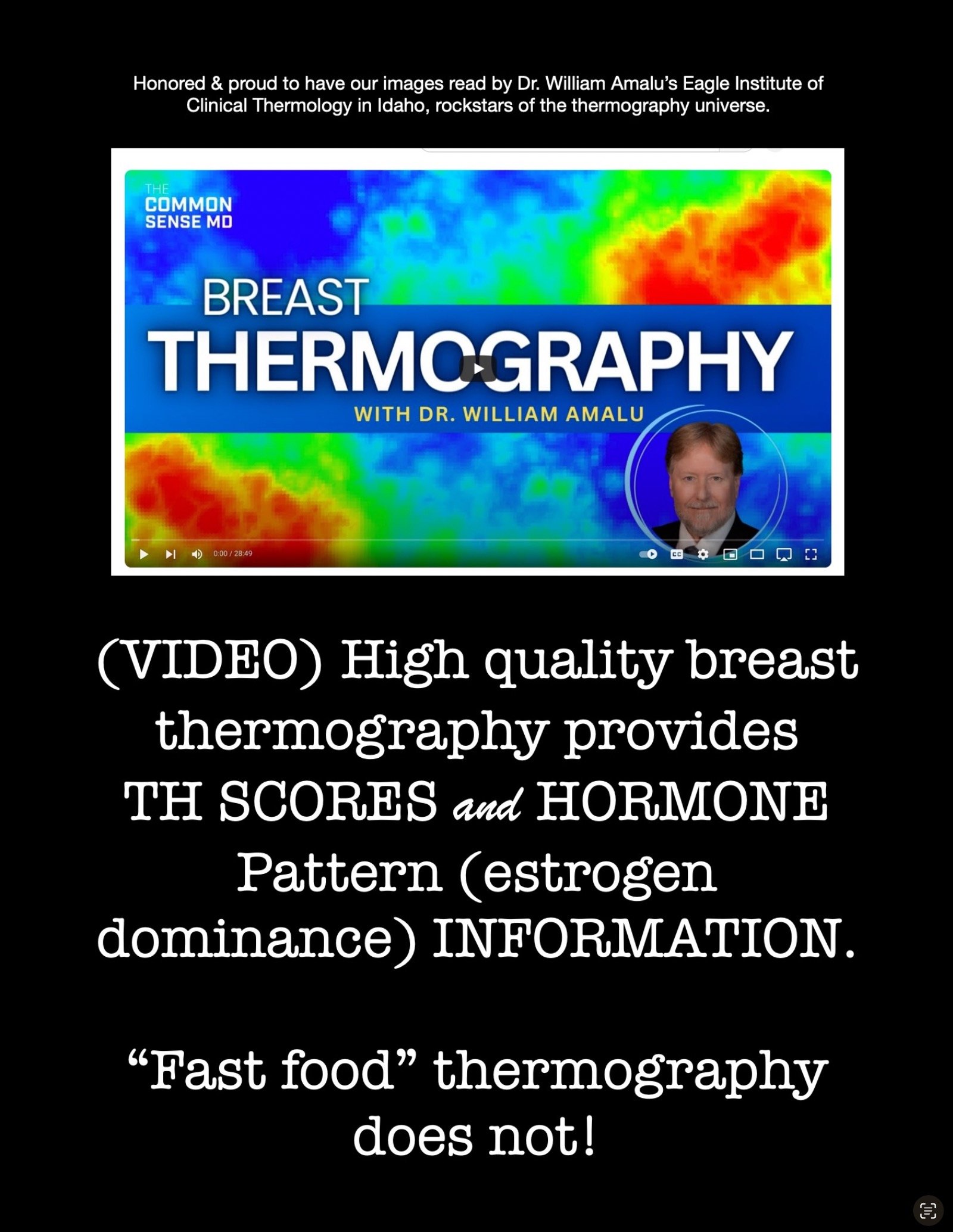
Click to watch video.
Excellent Interview above of Eagle Institute of Clinical Thermology’s Founder and Interpreter of our Images,
Dr. William “Bill” Amalu
This explains how quality thermography may be the first signal a woman receives that alerts her to estrogen dominance due to its ability to show patterns consistent with unopposed estrogen activity in breasts. Dr. Amalu also says estrogen in breasts can be up to 50 times the amount in blood, urine or saliva!
Estrogen Dominance, aka PROGESTERONE DEFICIENCY can hide in breasts while other tests (of blood, urine, saliva) may miss high estrogen activity in breasts.
Thermography is a true “canary in the coal mine” for early alerts about this dangerous estrogen excess so common in breasts today because of endocrine-disrupting “forever” chemicals that mimic estrogen and are called xenoestrogens.
Your hormone pattern status (i.e., not present, is present, better/worse etc) is given on each breast report of ours, so that if you have this pattern, you can watch your lifestyle changes take effect and/or be signaled if the pattern appears appears or worsens, true prevention strategy.
Occasionally, when Dr. Amalu is unavailable, his thoroughly trained with years of experience colleague, Dr. Valerie Quijano, interprets images for us/you. They are equally fabulous and accessible for questions you or your doctor may have.
What Will My Breast Thermogram Report Tell Me?
Dr. Amalu will analyze your images and prepare a detailed, easy to read report, noting any abnormalities if present.
Unlike 99% of our competition, we also take closeups of each breast and our reports indicate any areas of concern by the quadrant or specific “o’clock area” in which they occur that you may track over time. Each breast gets a separate numerical thermobiologic grade, not just a label of “at low risk” or “at some risk” or “at increased risk” as many other readers provide.
You will receive your images in high resolution grayscale as well as your choice of several color palettes from which to choose a set of color images too. A Guide will accompany your report explaining the different possible grades as well as explaining the thermal assessment for possible presence of hormone imbalance in your breasts. Your report will state either that you have this hormone imbalance pattern or that you do not and it will make recommendations about future imaging and possibly further testing.
Thermography, technology developed by NASA, offers you an early warning system that shows neurochemical signals far in advance of significant damage to the body. This means you and your health care providers can outline a treatment method for current or future problems before they cause irreversible damage. A sophisticated medical infrared camera and computer system scans the body surface for thermal (heat) abnormalities. Abnormal heat patterns are produced when dysfunctional areas send nervous system signals to the body’s surface.
Judy Mayer
Retired Mammographic & Thermographic Technician
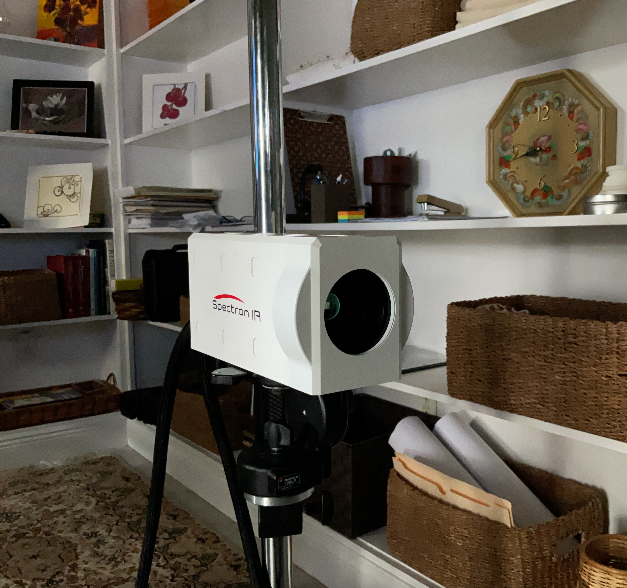
What Camera/Software Do We Use?
Our clinical infrared imaging system is cutting edge, designed for hospital use and cleared by the FDA for the greatest number of medical uses a camera can have. The software we use is TyTron C-500IR. Our SpectronIR cameras are cleared by the FDA for the highest uses in adjunctive diagnostic screening for the detection of breast cancer, neuromusculoskeletal disorders, vascular disease, metabolic, inflammatory and other neoplastic disorders. We also have a FLIR.
We use a secure, HIPAA-compliant, method of direct, safe transmission of your report and images to you.
Is Thermography a Substitute for Mammogram?
No. Is mammography a substitute for thermography? No. Thermography sees physiology in the form of heat. Mammography sees structure such as solid tumors. Ultrasound and MRI also see structure, and none of the structural modalities can see heat/physiology. In most cases, the aggressiveness of a tumor is in direct relationship to its thermovascularity (blood vessel activity). Benign lesions typically have no thermal markers. Infrared imaging offers a safe noninvasive procedure that is a valuable adjunct in determining whether a lesion is benign or malignant PRIOR to BIOPSY! We believe the negative biopsy rate could be improved dramatically if infrared imaging became standard of care prior to biopsy decisions. It improves their odds of being correct dramatically!
Thermography is the best risk-assessment tool known for predicting risk of breast cancer. Yet it is not covered by insurance. Hmmm. Did you ever question how often the name of your insurance company is the same name as that of your hospital. Is this not the greatest conflict of interest on the planet?
How to Avoid
“Fast Food” Junk Thermography & Get QUALITY!
THE QUALITY OF YOUR REPORT will make or break you, literally. FAST FOOD THERMOGRAPHY, like a diet of Halloween candy, will steal health while giving a wicked false sense of euphoria and security to those thinking all thermography reporting is equal. It’s similar to thinking all surgery is good ~ no, it hugely depends upon the surgical team.
As in cuisine, inferior “fast food” thermography is much more available than high quality imaging by about 20 to 1 in our country, according to Dr. Amalu, of Eagle Institute of Thermology and President of the International Academy of Clinical Thermology, and interpreter of our images.
QUESTIONS TO ASK
1
Are products being sold, for example, potions, lotions, latest gadget, etc., as “solutions” to “improve” health or thermal patterns (that never or rarely get better?) while pricing thermal imaging services way below the normal rates, as “loss-leaders”? This is snake oil selling at its darkest. Run from these imaging places or risk spending tons on worthless “treatment.”
2
Does the report indicate whether or not you have the presence of a very common pattern in today’s women that may indicate unopposed estrogen/progesterone deficiency, aka “estrogen dominance,” an imbalance DIRECTLY linked to the most common breast cancers, and does it recommend to see your provider for testing if you do have this pattern and alert you to possible causes to discuss with your doctor?
3
Does the report give precise locations on the breast by quadrant or “o’clock” position of any questionable heat patterns?
4
Does the report give a numerical TH (thermo-biological) Score for each breast?
5
Is the report signed by a doctor who also has the courage to put their phone number on the report & actually answer the phone?
With our reports, the answer is a huge “YES!” You receive all this and much more, yet none of it with fast-food thermography. We provide a report even your medical doctor can respect if educated enough to do their own research on PubMed and not trust uptodate.com because there is a literal conspiracy in place to repress and smear quality thermography.
(Fast-food) thermography reports are rightfully not respected by many doctors because they lack information and are truly dangerous. Real protection requires a real report with multiple specifics about each breast, information about the pattern suggestive of estrogen dominance and more, and that can stand up in court should the need ever arise.
YOU WILL GET DETAIL
with our reports that can improve your life instead of sabotaging it.
Physician confirmed estrogen dominance.
Our reports always let you know whether you have a pattern that may suggest hormonal imbalance and each subsequent report will let you know if the pattern has improved or gone away, etc. Fast-food thermography typically lacks this detailed analysis.
Estrogen a more powerful breast cancer culprit than we realized
says the medical system
The Harvard Gazette, May 17, 2023
Well well well, the same conglomerates who own hospitals and chemical companies that make glyphosate (a powerful xenoestrogen) want you to know that they have only just recently realized it is in fact estrogens not only fueling a huge percentage of breast tumors, but actually CAUSING them via “hot spots” @ estrogen receptor sites in breasts. Until now not admitted. Yet in the article they stop short of including xenoestrogens, only one of the big elephant endocrine disruptors in the room this century. So many truths are coming out as the medical authorities essentially reverse another fairytale they’ve told for decades in that article.
Headquarters Studio in Florida


Our headquarters imaging studio is in a neighborhood in Fort Walton Beach, one block from Uptown Station Shopping Center. Please park in the driveway if you like! 111 Opp Blvd NE, Fort Walton Beach, FL 32548.
Our Imaging Cottage in SE Portland Oregon

Our Portland, Oregon Clinic - a lovely little private hidden by bamboo cottage in southeast Portland, 4141 SE Crystal Springs Blvd #B, Portland, OR 97202
What is Breast Thermography & What Can It Detect?

Short answer is that it is a diagnostic procedure that images the breasts to aid in the early detection of breast cancer, is non-invasive, safe, uses no radiation, and is an effective means to help in the diagnosis or monitoring of:
Benign Breast Tumors
Breast Cancer
Breast Cancer Risk
Breast Mastitis
Fibrocystic Breast Disease
Breast Cancer Treatment
(click here for research and references)
Happy Healthy Breasts
Not Happy


This generous client was willing to share her images with you. Pictured is her right breast with visible blood supply feeding 3 cancerous tumors. We’ve since upgraded to Spectron cameras; these were taken with a FLIR A40.
GOT
Implants?
Dense Breasts?

Thermal imaging is not reduced in sensitivity nor is it negatively affected in any way by dense breasts or implants.
Pregnant or Nursing?
No Problem! But the “fast food” thermography clinics will not image you. We offer the skill necessary for reading your images even if you are pregnant or nursing.
Full Body Thermography
A sophisticated medical infrared camera and computer system scans the body surface for thermal (heat) abnormalities. Abnormal heat patterns are produced when dysfunctional areas send nervous system signals to the body’s surface.
(click here for research and references)
The scan is completely non-invasive, does not use radiation, and is a safe and effective means to help in the diagnosis of:
Arthritis
Diabetes
Headaches
Neck and Back Problems
Metabolic Disorders
Nervous System Disorders
Pain Syndromes
Repetitive Strain Injuries
Soft Tissue Injuries
Stroke Risk
TMJ Conditions
Vascular Disorders
And much more …
Click the link below for more information about what this technology can detect.
Over thirty years of clinical use and more than 8,000 peer-reviewed studies in the medical literature have established thermography as a safe and effective means to examine the human body.
Got Unresolved PAIN?
Thermography excels in aiding in the diagnosis of unresolved, elusive pains.

Watch this to understand the way the medical industry can twist statistics into what some would classify as “fear mongering” to motivate patients.
Stay Radiant
-
Screen Safely
-
Stay Radiant - Screen Safely -
“Canary in the Mine” for signaling ESTROGEN DOMINANCE a Heat Pattern in Breasts - True Prevention!
BIG IMPROVEMENTS
in Estrogen Dominance
(aka Progesterone Deficiency)
Pattern!

Two years and 15-17 hours a day of intermittent fasting brought “significant improvement” in this person’s estrogen dominance pattern!

Right breast’s BIG IMPROVEMENT in only 6 months of diet change.
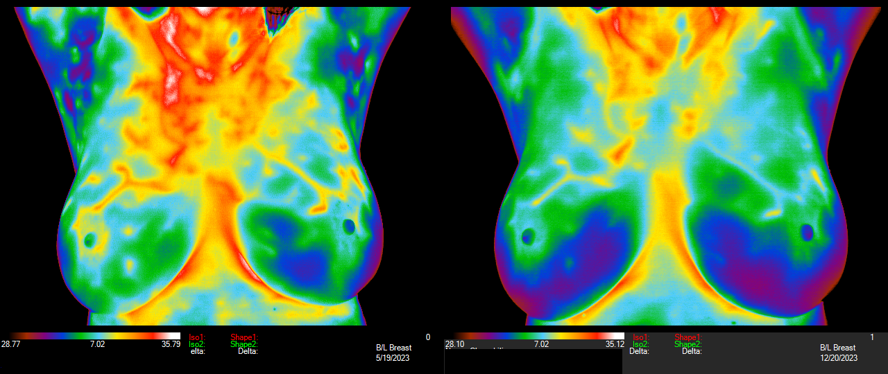
Six Months of Eliminating Soy and Flax! May to Dec 2023. Report states “significant improvement” in the pattern of hormone activity and her right breast went from a TH3+ to a TH3-. Beautiful!
Find out if you have the pattern suggestive of excess estrogen in your breasts & if so, consider changes to improve it, either with your provider or on your own, to lower your risk - proof is in the pictures!
For some, they and their practitioners find that diet and lifestyle changes will do it and others find they need to employ a hormone balancing cream containing progesterone formulated from only natural sources.

LAST YEAR ON THE LEFT - “SIGNIFICANT IMPROVEMENT” (REDUCTION) IN THE ESTROGEN DOMINANCE PATTERN - Sharon after 8 months of a transdermal progesterone cream on the right.

2022 August
Right Breast TH3-
Left Breast TH3+

2023 September
Right Breast TH1 (TH1’s are extremely rare today!)
Left Breast TH3
One client even found a sudden reduction in work/stress may have been the reason for her dramatic improvements, suggesting her hormones balanced when she had or put less demands on herself.
HOT Pattern in August 2022 images suggesting hormone Imbalance “RESOLVES”/Leaves by Sept 2023 and Both TH Scores Improve!!!
What did Judy do? Inquiring minds want to know so we asked!
Adios Abnormal!

Something WONDERFUL AND AMAZING happened in 2020! This Client reversed her TH4 ABNORMAL breast down to a TH3 (TH3 Grade means 95% chance of no pathology present) in 9 MONTHS! Click above to see more of her amazing images and learn how you too can be tipped off to EXCESS ESTROGEN, likely before it will show up in lab tests. William C. Amalu, DC, DABCT, says “thermography truly is the proverbial canary in the [coal] mine” to alert women far in advance to changes occurring that are conducive to cancer initiation and are catalysts of its promotion. These hot estrogenic vascular formation effects of glyphosate, soy, flax and other phytoestrogens & synthetic xenoestrogens have a heat signature seen with infrared imaging verified by MRI imaging.
If you use bio-identical hormones, glyphosate, soy, flax and any other phytoestrogens, it is especially important for you to receive thermography so that you know early if your breasts are “lighting up” due to possible excessive accumulation of estrogens in breast tissue since breasts can contain 50X as much estrogen as blood, saliva, or urine.
What doctors are saying about breast screening
(Our publication of the following doctors’ statements does not imply or suggest their endorsement of our service. We are happy to highlight their work and opinions, but this is not an endorsement of everything they say and do.)

Christiane Northrup, M.D.
Women’s Health Expert, Visionary Health Pioneer, Author
from The Best Breast Cancer Screening Tests: 6 Reasons Why I Recommend Thermography
from Thermography: A Much Saner and Less Invasive Approach
(especially for those carrying BRCA gene according to Northrup)

Michael Greger M.D.
Author NY Times Bestseller "How Not to Die"
www.nutritionfacts.org
50 videos/articles about Breast Cancer
"Breast cancer can take decades to develop, so "early" detection via mammogram may be too late.... "early detection" may in effect be really, really late detection."
Mushrooms for Breast Cancer Prevention

Thomas Hudson, M.D.
Author, Radiologist, Thermologist
Dr. Philip Getson, DO

Dr. Philip Getson, DO
Dr. Philip Getson, DO from The Best Breast Test: The Promise of Thermography

"Especially Valuable for Women with Dense Breasts"
American Journal of Surgery about Infrared Imaging:
In a study of the effectiveness of thermal imaging in detection of breast cancer, thermography identified “58 of 60 malignancies with 97% sensitivity” and was concluded to be “a valuable adjunct to mammography and ultrasound, especially in women with dense breast parenchyma."